Edema Disease

First hand experience

The disease is characterized by edema at specific sites, sudden unexpected death, and also by neurologic signs which are related to lesions in the brain.

While the Shigatoxin (Stx2e) encoding E. coli occur worldwide, prevalence data particularly from healthy herds are rarely available. Nevertheless, a recently published longitudinal study in three US farms with healthy pigs observed a high prevalence (68.3%) of Shigatoxin-encoding Escherichia coli (STEC) in pigs raised for pork production. The usual detection rate of Stx2e in cases with clinical signs suspicious for ED is around 20% up to 60%.

Pathogen

Escherichia coli is a bacterium that is commonly found in the lower intestine of animals. Nonpathogenic, intestinal Escherichia coli (commensal E. coli) support the physiological intestinal balance of the host, whereas pathogenic E. coli with typical virulence factor gene profiles can cause severe outbreaks of different diseases (e.g. Edema (Oedema) Disease, diarrhea).


 Edema (Oedema) Disease is caused by Shigatoxin (Stx2e)


Edema (Oedema) Disease is caused by Shigatoxin (Stx2e). This toxin is produced by a specific virotype of Escherichia coli – which is named STEC (graphic). A virotype is determined by a particular combination of virulence genes. Important virulence factors encoded by EDEC are the fimbrial adhesin F18 and Shigatoxin (Stx2e).
While the E. coli encoding for Shigatoxin (Stx2e) occur worldwide, prevalence data particularly from healthy herds are rarely available. Nevertheless, a recently published longitudinal study in three US farms with healthy pigs observed a high prevalence (68.3%) of Shigatoxin-encoding Escherichia coli (STEC) in pigs raised for pork production. The usual detection rate of Stx2e in cases with clinical signs suspicious for ED is around 20% up to 60%.

The Shigatoxin has an A and B subunit


Shigatoxins (Stx) are a family of related toxins with two major groups, Stx1 and Stx2. The toxins are named for Kiyoshi Shiga, who first described the bacterial origin of dysentery caused by Shigella dysenteriae. The most common sources for Shigatoxin are the bacteria S. dysenteriae and the shigatoxinogenic virotypes of Escherichia coli (STEC), and other enterohemorrhagic E. coli (EHEC).
The toxins are named for Kiyoshi Shiga, who first described the bacterial origin of dysentery caused by Shigella dysenteriae. The most common sources for Shigatoxin are the bacteria S. dysenteriae and the shigatoxigenic serotypes of Escherichia coli (STEC), and other enterohemorrhagic E. coli (EHEC).

Shiga-like toxins 1 and 2 are the Shigatoxins produced by some E. coli strains. Stx-1 is identical to Stx (true Shiga toxin – is produced by Shigella dysenteriae) or differs by only one amino acid. Stx-2 shares 56% sequence identity with Stx-1.

The name verocytotoxins/verotoxins – a seldom-used term for Stx – is from the hypersensitivity of the so-called Vero cells (used in cell cultures) to Stx. The toxin has two subunits—designated A and B (graphic). The B subunit is a pentamer that binds to specific glycolipids on the host cell. Following this, the A subunit is internalized and cleaved into two parts. The A1 component then binds to the ribosome, disrupting the protein synthesis. Stx-2 has been found to be about 400 times more toxic (as quantified by LD50 in mice) than Stx-1.
(Source: Sato et al. 2013 J. Vet. Med. Sci.; Tu et al. 2009 Protein Expression and Purification)

Mainly affected age group by Shigatoxin


The mainly affected production stages are recently weaned pigs, pigs during nursery and younger finishing pigs.
F18 is one of the well known adhesion factors, which enables E. coli the attachment to the intestinal mucosa. Stx2e encoding strains may possess either the fimbrial variant F18ab or F18ac.
Susceptibility to colonization and to infection with an E. coli F18 strain develops around weaning and is dependent on the presence of the porcine intestinal F18 receptor (F18R) genotype.
The expression of F18R is age-dependent and begins at around three weeks of age.
Torrison at al. (graphic) described the main stages of pigs life where E. coli encoding for Stx2e are detected with a peak in the weeks seven to nine. This corresponds with the experience of IDT in many countries worldwide.
Remarkably Stx2e can be detected also in later production stages . If so, it is generally the origin of serious clinical signs, increased mortality and impaired performance.

Yet F18 is not detectable in every case of E. coli encoding for Stx2e (up to 20%). The relevance of these particular E. coli (without the F18 but with Stx2e) is nevertheless given, as they have been isolated as only pathogen from cases with severe clinical signs.

J.Torrison, K. Rossow, S. Oliveira
University of Minnesota Veterinary Diagnostic Laboratory 2011


Edema (Oedema) Disease

Pathogenesis of Edema (Oedema) Disease (ED)

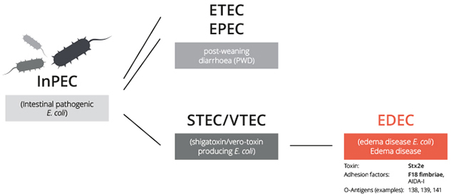
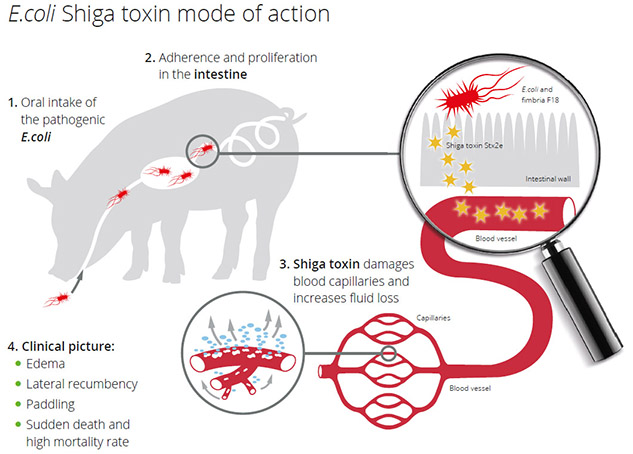
The causative agent of ED is Shigatoxin-2e. It´s encoded and produced by a specific virotype Escherichia coli and occurs worldwide in diseased pig herds as well as in healthy herds. The primary habitat E. coli in pigs is the gastrointestinal tract. Outside the intestine E. coli are found in fecal-contaminated feed or water, water from water pipes contaminated by biofilm (Wöchtl B. et al, Tierärztl). Umschau 72, 2017), soil, and the environment of the pig barn. The infection happens via the oral route. When ingested in sufficient numbers, E. coli causing ED colonize and then proliferate rapidly to attain massive numbers. Colonization develops over 3-6 days and requires attachment of fimbrial adhesins to complementary receptors on the small intestinal epithelium or in the mucus coating in the distal jejunum and ileum. Stx2e is absorbed into the circulation and causes vascular damage in target organs.

The degenerative angiopathy of small arteries and arterioles results in an increase in vascular permeability. Due to osmosis, fluid leaves the vascular system and diffundates into various tissues, causing lesions that can be seen macroscopically as edema.
Gelatinous edema appears in the submucosa of the gastric cardia and occasionally in the fundus, in the mesocolon, in the small intestinal mesentery and gallbladder bed. The mesenteric and colic lymph nodes may be swollen, edematous and congested. Edematous swelling of eyelids, forehead, larynx and brain can be observed.
(Source: Diseases of Swine, Tenth Edition, Edited by J.J. Zimmerman et al., John Wiley & Sons, Inc. Published 2012)

Clinical signs of Edema (Oedema) Disease (ED)

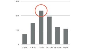
ED mostly occurs in recently weaned pigs, although cases may be observed in later production stages as well. The disease may be sporadic and may affect only individual animals, but occasionally an entire batch of pigs is affected.

The subacute/ acute outbreak often is recognized as sudden death without previous signs of sickness. Pigs dead of ED are mostly in good condition. Some affected pigs become anorexic, develop swelling of the eyelids and forehead. As a result of the laryngeal edema some pigs emit a peculiar squeal. Due to the edema in the brain pigs show convulsions, ataxia and lateral recumbency with paddling of limbs. Few pigs survive the acute disease, but those that do remain runts (i.e. show stunted growth).

The course of the disease can also be prolonged. Clinical signs then reoccur in the same batch of pigs, especially after further change of feed, removal of zinc oxide (ZnO) or antimicrobials from the feed or another stress factor.

Subclinical ED may also occur, where pigs are clinically normal but develop microvascular lesions and may have a decreased growth rate. These herds may show inhomogeneous groups of pigs in the different production stages.

Chronic ED occurs in a low proportion of pigs recovering from acute disease or in herds where chimeric E.coli, which produce the heat labile (LT) and heat stabile (ST) toxins that cause post-weaning diarrhea (PWD) but also produce Shigatoxin-2e, are detected. For periods varying from days to several weeks after intestinal infection, growth stops and sick pigs often show unilateral nervous disturbances such as circling movements, twisting of the head, or atrophy of limb muscles with progressive weakness. In these cases subcutaneous edema is rare.
(Source: Diseases of Swine, Tenth Edition, Edited by J.J. Zimmerman et al., John Wiley & Sons, Inc. Published 2012)