Neonatal Diarrhoea
Importance and economical relevance of neonatal diarrhoea
Neonatal diarrhoea is one of the most common disease conditions on a pig farm today. In recent years, pig production has changed and intensification has increased, consequently the number of animals on the farm is increasing, with associated changes in management. The intensive farming contributed to the increase in occurrence of neonatal diarrhoea which causes significant economic losses and excessive use of antimicrobials in the farrowing house.
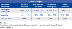
Estimated costs for herds affected by neonatal diarrhoea with mortality of 10 % caused by disease can be as high as 134 Euro per sow per year. Together with Swine dysentery, neonatal diarrhoea belongs to the most expensive enteric issues on pig farms (tab. 1 and fig. 1) (Sjolund et al., 2014).
The frequency of neonatal diarrhoea cases increased in the EU in recent years affecting classical farms with conventional herd health status as well as well-maintained farms with good management practices.
Pathogens of the neonatal diarrhoea complex
Neonatal piglet diarrhoea is a very common and relevant problem in modern pig production. It is associated with increased pre-weaning mortality, poor growth rates and variation in weight at weaning. The newborn pig has an immature mucosal immune system at birth allowing pathogens to colonize the gastrointestinal tract immediately after birth.
By definition, neonatal diarrhoea is characterised by diarrhoea that develops during the first week of the piglet‘s life (usually within the first few days after birth).
Escherichia coli complex
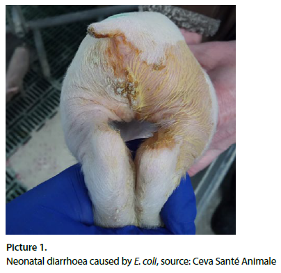
Regarding bacterial infections, Escherichia coli (E. coli) has historically been considered one of the main agents causing neonatal diarrhoea in pigs (Chan et al., 2013). Different E. coli pathotypes have been identified based on toxin production and other virulence factors. The most common are the enterotoxigenic E. coli (ETEC) strains, characterized by the production of enterotoxins (STa, STb and LT) and different fimbriae (adhesins). Both – toxins and fimbriae are considered as virulence factors. Other pathotypes of E. coli have been detected in piglets, such as enteropathogenic E. coli (EPEC) strains, producing intimin (eae gene), although less frequently and its practical importance is rather low (Toledo et al., 2012). ETEC responsible for neonatal diarrhoea possess adhesins, surface proteins called fimbriae, identified as F4 (k88), F5 (k99), F6 (987P) and F41. The fimbriae allow the microorganism to adhere to specific receptors on the brush borders of the small intestines’ enterocytes.
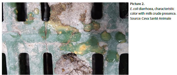
The most prevalent ETEC with the fimbriae F4 colonize the length of jejunum and ileum, while ETEC with fimbriae F5, F6, F41 mostly colonize the posterior jejunum and ileum. Susceptibility to ETEC F5, F6 and F41 decreases with age and has been related to a reduction in the number of active receptors present on the intestinal epithelial cells. The most ETEC strains of neonatal colibacillosis produce heat stable enterotoxin STa, which binds guanylyl cyclase C glycoprotein receptor on the brush border of villous and crypt intestinal epithelial cells, stimulating the production of cyclic guanosine monophosphate (cGMP) leading to electrolyte end fluid secretion and consequent watery diarrhoea if the excess fluid from the small intestine is not absorbed in the large intestine. Excessive secretion leads to dehydration, metabolic acidosis, and eventually death (Diseases of Swine, 10th edition).
Clostridium complex
Anaerobic bacterial pathogens such as enterotoxigenic strains of Clostridium perfringens type A (CpA) (producing Cpα toxin, β2 toxin), C. perfringens type C ( Cpβ toxins) are important pathogens in the complex of neonatal diarrhoea and diarrhoea before weaning (Uzal and Songer, 2019). Clostridium difficile (C. difficile) producing enterotoxin A (TcdA) and/or cytotoxin B (TcdB) can be as well isolated and can cause similar disease to CpA. Clostridia are large, sporulating, gram-positive rods. Pathogenic clostridia produce one or more toxins with direct or indirect evidence links with pathogenesis in neonatal diarrhoea. The widely used system of classification of C. perfringens is based on presence of Major toxins gene and toxin production on five toxin types: α, β, ε, τ, θ. From swine medicine perspective CpA and CpC are dominant pathogens, causing infection of piglets.
Clostridium perfringens type C
CpC is a primary pathogen of piglets. Organisms persist in the environment and are resistant to heat, disinfectants and UV radiation. Multiplication of CpC in affected piglets after infection from mother or from contaminated environment causes haemorrhagic necrotic enteritis, which is characterised by bloody diarrhoea, accompanied by high mortality. The case fatality is up to 100 %. Infection can be as early as 12 h after birth, usually within the first 7 days of age (DOA). Sows are the source of infection to piglets. Newborn pigs are able to acquire passive systemic immunity by absorption of undigested immunoglobulins from colostrum during the first 24-36 h after onset of colostrum ingestion. Colostrum trypsin inhibitors were suggested to be of possible pathogenetical significance in enteritis in newborns. The β-toxin of CpC is inactivated by trypsin and could easily be protected by the trypsin inhibitor in sow colostrum (P.T. Jensen, https://hal.archives-ouvertes.fr/hal-00900993, 1978). Infection occurs epidemically on farms where effective vaccination is not applied. Hallmark lesions are profound, usually segmental, mucosal necrosis with marked hemorrhage and emphysema of small intestine, principally the beta1 toxin acts systemically as well.
Clostridium perfringens type A
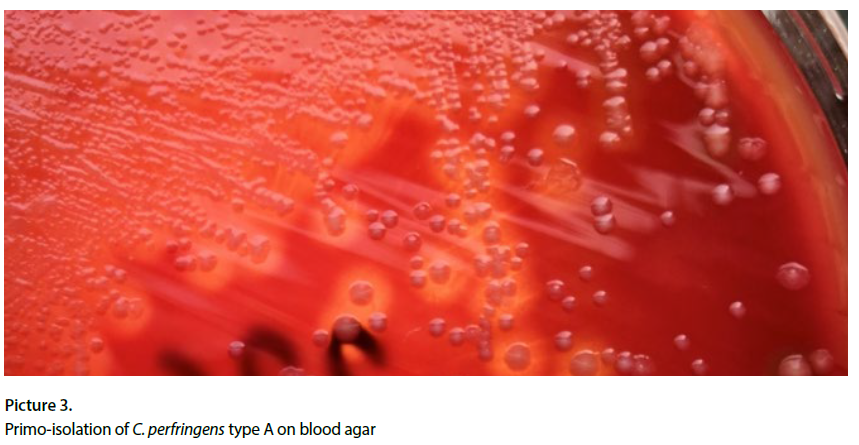
CpA is included in the microbiota of the swine intestine and properly equipped strains are able to cause enteric disease. Infection of piglets is characterised by mild inflammation of the mucosa, occasionally with adherent necrotic material. Microscopic lesions may include superficial damage of epithelial tips of the villi – primarily localized in the jejunum and Ileum. Macroscopic changes of intestinal mucosa might be subtle and discrete. In such cases microscopic assessment would be needed. Diarrhoeic cases usually correspond with P-A affection of intestine and presence of large amount of pathogenic bacteria associated with lesions.
Principal toxin type is alpha toxin, together with beta2 toxin (CPB2; cpb2 is the encoding gene, Jost et al., 2005). For the production of toxin, adherence of the pathogen to the mucosa plays a role. Clinical signs were reproduced experimentally (Johannsen et al., 1993). Infection is characteristic for the first days (one week) of life of the piglet and the source is usually the sow. CpA is as well a very early colonizer of piglets after birth. CPB2 positivity of CpA (PCR detection) is used as differentiation between normal flora and pathogenic type A strains, causing disease. Diarrhoea has a creamy or pasty character, lasts usually for up to 5 days and presence of mucus but without blood is frequent. Inflammation of mucosa is mild compared to CpC, at necropsy, small intestine is flaccid, thin walled and gas filled with watery contents.